

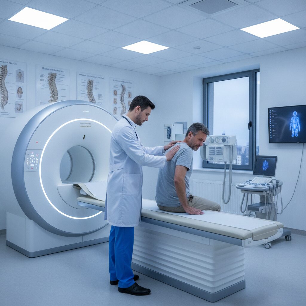
Most people with leg pain that starts in the lower back never need a scan. The pain flares, they adjust how they sit and move, get conservative care, and things settle over a few weeks. But a smaller group of patients walks into the office with a story that does not fit that pattern, and for them, going straight into treatment without a clear picture of what is happening inside the spine can waste time or even cause harm. That is why any thoughtful approach to sciatica treatment Rochester Hills residents can trust starts with a simple question: does this case need imaging first?
Imaging is not a magic key, and it is not a box to check. An MRI or X-ray can reveal things a hands-on exam cannot, but it can also show age-related changes that have nothing to do with your pain. The skill is in knowing when a picture will change the plan and when it will only add cost and anxiety.
In this article, we will walk through the situations where imaging earns its place, the red flags that move a case to the front of the line, the difference between the main scan types, and how to talk with your provider about it. Whether you run a small business and cannot afford weeks of downtime, spend your days at a keyboard, or simply want straight answers, this guide will help you ask better questions and make calmer decisions.
Most Sciatica Does Not Start With a Scan
It surprises many people to learn that the majority of sciatic pain episodes improve without any imaging at all. The sciatic nerve is the largest nerve in the body, running from the lower back through the buttock and down the leg, and when it gets irritated, the pain can be sharp, burning, or electric. In a typical case, the cause is a mechanical irritation near the lower spine, and a careful history plus a physical exam gives enough information to start safe, conservative care.
This is the same logic behind a good Rochester Hills sciatica treatment clinic intake: listen first, test the nerves and joints, and watch how the body responds in the first few visits. If symptoms steadily improve, a scan would rarely change what happens next. Ordering one anyway can show disc bulges or degeneration that are common even in people with no pain, which can make a manageable problem feel scarier than it is.
So imaging is not the default. It is a tool reserved for cases where the exam, the timeline, or the symptoms suggest something more is going on.
Red Flags That Move a Case to the Front of the Line
Some symptoms tell a provider that waiting and watching is not the right call. These are often called red flags, and they point toward conditions that need to be ruled out quickly. The most urgent include new loss of bladder or bowel control, numbness in the groin or inner thighs, rapidly worsening leg weakness, and severe pain that does not ease in any position. Any of these should prompt same-day medical attention, not a routine appointment.
Other warning signs are less dramatic but still matter. Unexplained weight loss, fever alongside back pain, a history of cancer, or pain that wakes you reliably at night can suggest causes beyond a simple irritated nerve. A recent fall, car accident, or other trauma is another reason to look before treating, because a fracture or instability would change which techniques are safe.
Think of red flags as a filter. Most people pass through without a single one, and conservative care proceeds. The few who trigger a flag get a faster, more careful path, which is exactly how the system should work.
When Time Is the Deciding Factor
Even without red flags, the calendar can change the conversation. If leg pain has been steady for roughly four to six weeks despite sensible care, many providers will consider imaging to see whether a structural problem is blocking progress. The exact window varies by person and situation, and it is a guideline rather than a hard rule, but the principle is sound: a case that is not moving deserves a second look.
The opposite is also true. A case that improves a little each week, even slowly, usually does not need a scan. Improvement in range of motion, walking tolerance, or the distance the pain travels down the leg is a strong sign the current plan is working. Providers often track these details because they say more about progress than any picture could.
If you are self-employed or work in a physical job, it is tempting to push for a scan early just to get an answer. Understandable, but patience paired with honest tracking tends to serve people better. Bring a simple log of your pain levels and activities to each visit so the timeline is visible.
What an Exam Can and Cannot Tell You
A thorough physical exam reveals a lot. Reflex tests, muscle strength checks, sensation mapping, and movement screens such as the straight leg raise can point toward which nerve root is irritated and how severely. A skilled Chiropractor in Rochester Hills, MI will also assess the hips, pelvis, and surrounding muscles, since these often contribute to nerve irritation in ways a scan would not show.
What an exam cannot do is look directly at bone, disc material, or the narrow channels the nerves travel through. If the findings are inconsistent, such as pain that does not match any single nerve pattern, or if the exam raises suspicion of fracture, tumor, or significant stenosis, imaging fills in what hands cannot feel.
The best practice treats exam and imaging as partners rather than rivals. The exam decides whether a picture is needed, and the picture, when ordered, is interpreted in light of what the exam found. A scan result that does not match your symptoms is often set aside rather than chased.
Choosing Between X-Ray, MRI, and Other Studies
Not all imaging answers the same question, so the choice of study matters. X-rays show bone structure well, making them useful for fractures, alignment, arthritis, and spinal curvature, but they cannot show nerves or discs directly. MRI uses magnetic fields rather than radiation and displays soft tissue in detail, including discs, nerve roots, and the spinal canal. CT scans offer sharp bone detail and are sometimes used when MRI is not an option. Nerve conduction studies are a different category, measuring how well electrical signals travel along a nerve.
Here is a quick comparison to keep the options straight:
| Study Type | Best At Showing | Common Reason It Is Ordered |
|---|---|---|
| X-ray | Bone alignment, fractures, joint wear | Trauma, suspected instability, structural concerns |
| MRI | Discs, nerve roots, spinal canal | Persistent leg pain, weakness, or numbness |
| CT scan | Detailed bone structure | When MRI cannot be used |
| Nerve conduction study | Electrical function of nerves | Unclear nerve involvement after exam |
Why Scans Can Sometimes Mislead
One of the hardest ideas to accept is that a scan can look alarming in someone who feels fine. Studies of people without back pain routinely find disc bulges, degeneration, and other changes that are simply part of aging, much like gray hair or wrinkles. When these findings show up in a report, they can sound frightening, yet they may have no connection to the pain you are feeling.
The reverse also happens. A scan can come back looking quite normal while a person is in real discomfort, because nerve irritation, muscle tension, and joint dysfunction do not always leave a visible mark. This is why providers lean on the whole picture rather than any single result.
There is a psychological side too. People who see a dramatic report sometimes become fearful of movement, avoiding the very activity that would help them recover. A good provider explains findings in plain language and keeps the focus on function: what you can do, what you cannot, and what is changing week to week.
Hidden Contributors a Picture May Not Reveal
Because sciatica is a symptom rather than a diagnosis, its sources can be varied. Tight hip rotators, a restricted sacroiliac joint, or a tender muscle in the buttock can all mimic or amplify nerve pain without ever showing up on an MRI. In these cases, imaging may look unremarkable while the real driver is movement and muscle behavior.
Functional testing helps here. Providers may watch how you stand, walk, bend, and transfer weight, looking for compensation patterns that load the lower back unevenly. Some offices also use muscle testing methods to guide care, a topic explored in Understanding Applied Kinesiology in Rochester Hills Chiropractic Offices, which can complement a standard exam when the picture is not clear.
The takeaway is that a normal scan does not close the case, and an abnormal scan does not automatically explain it. Imaging is one data point among several, and the plan should reflect all of them.
Everyday Habits That Can Muddy the Picture
Daily routines can quietly shape both symptoms and what a provider finds. Hours at a desk, a worn-out work chair, or a car commute can load the lower spine in ways that keep nerve irritation alive. That is why a history that includes your workday matters as much as the scan itself, and why reading about How Poor Posture Contributes to Back Pain in Rochester Hills can give useful context before an appointment.
If you are a developer, a shop owner on your feet all day, or someone who drives for work, tell your provider specifically what your day looks like. Details such as how long you sit before standing, what your monitor height is, and whether pain worsens at certain times can steer the plan more efficiently than a general description.
Small adjustments, such as a short walk every hour or a supportive seat cushion, are low risk and often worth trying while the evaluation continues. They will not replace a proper workup when one is needed, but they can ease the load on an irritated nerve in the meantime.
Finding the Right Local Care in Rochester Hills
When you are searching for care in Rochester Hills, it helps to look for a provider who explains their decision-making rather than ordering tests by reflex. Ask how they decide when imaging is needed, whether they coordinate with medical doctors or radiologists when a case calls for it, and how they will track your progress. A practice that is comfortable saying “we do not need a scan yet, and here is what would change that” is usually one that puts the patient first.
It is also reasonable to ask about referral pathways. If something concerning shows up, you want a team that knows exactly where to send you and how quickly. That coordination is part of safe care, and it should be spelled out before you need it.
Finally, trust your sense of whether you are being heard. Good care feels collaborative. You should leave visits understanding what was found, what the plan is, and what signs would prompt a change in direction.
How to Prepare for the Conversation About Imaging
A little preparation makes the imaging discussion far more productive. Before your visit, jot down when the pain started, what makes it better or worse, whether it travels below the knee, and whether you have noticed any numbness, tingling, or weakness. Note any past injuries, surgeries, or imaging you have had, and bring copies of earlier reports if you have them, since comparisons are valuable.
Here are a few questions worth asking:
- What do you think is causing my symptoms, based on today’s exam?
- Would imaging change the treatment plan right now?
- What signs would mean I should get a scan sooner?
- If a scan is ordered, what type, and why that one?
- How will we know the care is working?
Questions like these signal that you want to be a partner in decisions, and they tend to draw clearer, more practical answers.
What to Expect After the Scan Results Arrive
Once results are in, the next step is interpretation, not panic. A report may use technical phrases like protrusion, stenosis, or degenerative change, and your provider should translate these into what they mean for you specifically. Ask whether a finding lines up with your symptoms, because matching findings carry far more weight than incidental ones.
For many people, the results simply confirm that conservative care is appropriate and add helpful detail about which movements or positions to avoid. For others, the scan reveals something that calls for a medical referral, and having found it early is exactly the benefit imaging is meant to deliver.
Either way, the plan should be updated rather than put on autopilot. Expect a clear next step, a timeline for reassessing, and honest guidance on activity. Results are a starting point for decisions, not a verdict on your future.
The Bottom Line
Imaging in sciatica is about judgment, not routine. Most cases improve with conservative care and never need a scan, while a smaller group benefits greatly from one because of red flags, a stalled recovery, trauma, or an exam that does not add up. Knowing which group you fall into is the whole game, and the answer comes from a careful history and hands-on evaluation rather than from the machine itself.
Keep a few principles in mind. Urgent symptoms such as loss of bladder or bowel control, groin numbness, or rapidly worsening weakness need immediate medical attention. A case that has not budged after several weeks deserves a second look. Scan findings should always be read alongside your symptoms, since many changes seen on images are common in people without pain. And everyday factors like posture, work setup, and movement habits still matter whether or not a picture is taken.
If you are dealing with leg pain that started in your back, the most useful step is a thoughtful evaluation with a provider who explains their reasoning and is willing to refer when needed. Track your symptoms, ask direct questions, and do not be pressured into unnecessary testing or talked out of necessary testing. With the right approach, you can match the level of investigation to the actual problem, save time and money, and get back to the work and activities that matter to you.